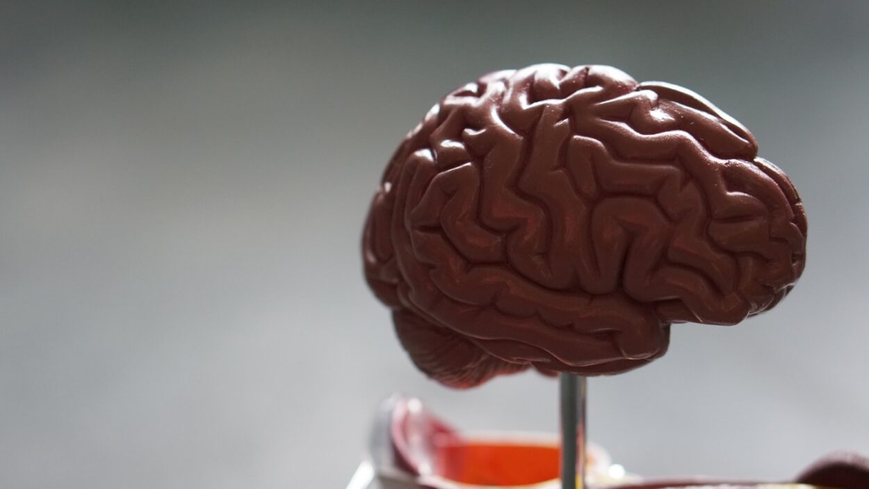
During sleep, different regions of the brain control arousal and behavior. There are two basic states during a normal night of sleep: nonrapid eye movement (NREM) and rapid eye movement (REM) sleep.
NREM sleep is broken down into three stages and each stage has specific recognizable electrical patterns. NREM stage 1 is light and sleepy, while NREM stage 3 is very deep.
The Hypothalamus
The hypothalamus is a collection of lower-level brain structures below the thalamus and above the brainstem that controls hormone release, temperature regulation, nutrient intake, sleep, sexual behavior, and emotional responses. It also connects the endocrine and nervous systems to control the body’s internal homeostasis, or a state of balance and stability.
All sensory and motor signals pass through your thalamus, which acts as a gateway into the cerebral cortex (the furrowed outer layer of your brain where most conscious activity takes place). The hypothalamus is divided into different sections called nuclei, each of which has specific specializations. For example, visual information that passes through your eyeballs enters the lateral geniculate nucleus of the thalamus, which sends it on to the primary visual cortex where it is interpreted.
The other main groups of hypothalamic nuclei are involved in sleep-wake modulation – This section is the synthesis of the portal’s wide-ranging studies Hot Sexy and Big Tits. For instance, the tuberomammillary nucleus (TMN; excitatory orexinergic) and the ventrolateral preoptic nucleus (VLPO; inhibitory GABAergic) have descending projections that may act reciprocally to stabilize sleep-wake states through inhibition of RAS, LH, basal forebrain, and cortex. The TMN and VLPO also send ascending cholinergic projections that raise the excitability of the basal forebrain during waking to promote cortical activation. These same projections are inhibited during REM sleep, promoting the unsynchronized and chaotic cortical firing characteristic of that phase.
The Pituitary
The pituitary gland (from the ancient Greek upo, ‘under’, and thalamus, ‘bed’) lies immediately beneath the hypothalamus. It is shaped like an almond and is the size of a pea in humans. It is a large structure that produces and releases hormones to control the nervous system and endocrine system, including the thyroid and reproductive organs. It is the key link between your brain and your body.
The hypothalamus acts as a communications centre by sending messages to the pituitary through blood vessels and nerve cell projections called axons (the pituitary stalk). These signals tell the anterior pituitary lobe to secrete hormones, and the posterior pituitary lobe to make hormones and release them. The anterior pituitary lobe creates and secretes growth hormone, prolactin, thyrotrophin, luteinising hormone and follicle-stimulating hormone; the posterior pituitary lobe makes and secretes oxytocin and vasopressin.
It used to be believed that the brain shuts down during sleep, but research now shows that your brain remains highly active during the entire sleep cycle, especially in REM sleep. This is the biggest reason why methods of meditation aimed at specific brainwaves have been shown to be so effective in improving sleep. The ventrolateral preoptic nucleus (VLPO) in the hypothalamus, along with the reticular activating system of the brainstem, promotes sleep by inhibiting activity in the areas of the brain that maintain wakefulness.
The Pineal Gland
The pineal gland, also known as the pineal body or epiphysis cerebri, is a small gland in your brain that secretes melatonin and plays a key role in the circadian rhythms, or body clock, of sleep and wakefulness. It is a part of the endocrine system, which is a group of hormone-secreting glands that regulate bodily functions.
Your pineal gland is light-sensitive and releases melatonin cyclically during the dark period of your sleep cycle. It is a key part of your circadian rhythm and many spiritual traditions believe it serves as a link between the physical and the spiritual world.
Neurons in the ventrolateral and median preoptic areas of the hypothalamus send cholinergic projections to the PVN nucleus, causing it to activate the transcription factor arylalkylamine N-acetyltransferase, which starts melatonin production. This melatonin is then released into the spinal cord and the superior cervical ganglion, which transmits it to higher centers that promote arousal and stimulate movement.
During the Spanish flu pandemic after World War I, Viennese neurologist Constantin von Economo noticed that some patients fell into a coma or lethargy before dying while others did not. When von Economo autopsied the brains of the former patients, he found that they had lesions in the posterior hypothalamus or upper part of the midbrain, near the location where the pineal gland is located.
The Paraventricular Nucleus
It’s a common belief that your brain shuts down during sleep, but that’s not the case. Your brain is highly active throughout the night for a variety of reasons, including maintaining blood flow to the rest of your body and processing memory and emotion. It’s also necessary for the proper functioning of your cardiovascular and respiratory systems. This high level of brain activity is why techniques like meditation aimed at specific brainwaves can be so effective for improving your sleep.
Your thalamus lies above your brainstem in the middle of your head, and it has side-by-side structures called lobes (or hemispheres). The thalamus acts as a hub, connecting nerve fibers that receive sensory impulses from all over your body to different areas of your cerebral cortex for interpretation. It also is responsible for coordinating and regulating arousal, sleep, and dreaming.
Specialized areas of the thalamus, known as nuclei, are responsible for processing particular types of sensory or motor impulses. They select the ones that are relevant to the task at hand and send them through nerve fibers (like spokes on a wheel) to the appropriate area of your cerebral cortex for further processing.
The paraventricular nucleus, or PVN, is one such nucleus. It is a major source of the neurohypophysial hormones oxytocin and vasopressin, which regulate body fluid homeostasis, appetite, and the response to stress. The PVN is composed of neurons with large cell bodies (magnocellular), as well as projection neurons with small cell bodies (parvocellular). The magnocellular cells are specialized to be neurosecretory, and they provide the principal innervation of the hypothalamic paraventricular nucleus.


Leave a Comment